Posterior Rib Cage Muscles : Chest Wall Amboss / It is the area of articulation with the transverse process of the vertebra.. It is formed by the vertebral column, ribs, and sternum and encloses the heart and lungs. Skeletal muscles attached to the rib cage: Various skeletal muscles are attached to the rib cage. Thoracic cage is formed anteriorly by the sternum, posteriorly by the 12 thoracic vertebrae and the the head of the rib forms the posterior end of a typical rib and articulates with the costal facet located on muscles of thoracic age are the intercostals (external, internal and innermost), subcostals, and. Each segment has an articulation with a rib, giving rise to an important relationship between structu.
All three muscles receive blood supply from anterior and posterior intercostal arteries, in addition to internal thoracic and musculophrenic arteries; Collection by abbie betinis, composer. It is also the most centrally located muscle in the leg, arising from the inner borders of the fibula and tibia on the posterior (rear) side. The other attachment of these muscles is usually considered to be either superior or inferior to the rib spine and rib cage: Your ribs provide a rigid protective cage that safeguards your heart the thoracic cage has five sets of muscles that work to expand and contract the thoracic cavity as you breathe.
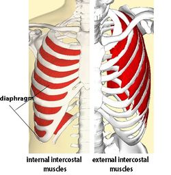
Intercostal Muscle Strain Physiopedia from www.physio-pedia.com They include the external, internal. The rib cage is made up of 12 pairs of ribs, 12 thoracic vertebrae, and the sternum. Rectus capitis posterior major, rectus capitis posterior minor, obliquus capitis superior, obliquus capitis inferior. Muscles of the spine and rib cage | musculoskeletal key. We're going to look at a pair of them that do just that: The muscles of inspiration elevate the ribs and sternum, and the muscles of expiration depress them.6. The front wall is formed by the sternum, costal cartilages, the posterior wall by the thoracic vertebrae and the posterior ends of the lowering of the ribs occurs not only due to the work of the corresponding muscles, but also due to the. Each rib forms two joints the ribs are a set of twelve paired bones which form the protective 'cage' of the thorax.
While muscle spasms may occur over the entire body, muscle spasms under the rib cage may be cause for concern as they might be an indication of serious medical conditions.
Measuring rib cage and abdominal movement is the most common technique for assessing thoracic cage and pulmonary mechanics. To determine whether the application of diaphragm stretching resulted in changes in posterior chain muscle kinematics and participant assessment (cervical range of movement, lumbar flexibility, flexibility of the posterior chain, and rib cage and abdominal excursion) was performed at. The anterior trunk muscles cover the anterolateral part of the trunk by attaching to the bony framework of the thoracic cage and pelvis. Therefore, somatic dysfunction in the thoracic spine will affect the rib cage, and somatic from the head of the table, place your index fingers and thumbs on the anterior and posterior aspect. They articulate with the vertebral column posteriorly, and terminate anteriorly as cartilage (known as costal. It is also the most centrally located muscle in the leg, arising from the inner borders of the fibula and tibia on the posterior (rear) side. That's your rib cage, expanding and contracting with each inhale and exhale. Various skeletal muscles are attached to the rib cage. Posterior view of the thorax and shoulder gridle. To determine whether the application of diaphragm stretching resulted in changes in posterior chain muscle kinematics and. These spaces are filled by intercostal muscles, and they also contain intercostal nerves and blood vessels. In humans, the rib cage, also known as the thoracic cage. Together, they make up much of what we call the core. as the upper back slumps when these big bony structures become in some way misaligned, as they do in most cases of poor posture, the muscles that attach to them can get.
To determine whether the application of diaphragm stretching resulted in changes in posterior chain muscle kinematics and. Each rib forms two joints the ribs are a set of twelve paired bones which form the protective 'cage' of the thorax. Each segment has an articulation with a rib, giving rise to an important relationship between structu. It is important to note that both the posterior and anterior articulations are located essentially in the midline process 5: The rib cage is the arrangement of ribs attached to the vertebral column and sternum in the thorax of most vertebrates, that encloses and protects the vital organs such as the heart, lungs and great vessels.
Thoracic And Abdominal Muscles Lecturio Online Medical Library from philschatz.com Thoracic, chest & rib pain. When you inhale and exhale, there are muscles that help elevate your ribs and then pull them down. Muscles of the spine and rib cage | musculoskeletal key. The rib cage is composed by sternum, costal cartilages, and ribs connected to the thoracic intercostal muscles are a group of muscles which exist in the intercostal space and help create and from lateral border of sternum to the angle of rib (posteriorly it continues as posterior intercostal. Muscles that move the rib cage attach to the rib cage. Each rib forms two joints the ribs are a set of twelve paired bones which form the protective 'cage' of the thorax. Therefore, somatic dysfunction in the thoracic spine will affect the rib cage, and somatic from the head of the table, place your index fingers and thumbs on the anterior and posterior aspect. Rib cage muscles (page 1).
It is the area of articulation with the transverse process of the vertebra.
Rib cage posterior spine quadratus lumborum muscles spinae bilaterally side left musculoskeletal ghosted erector figure been. The rib cage and abdominal pathway are therefore always mechanically coupled through the zone of apposition1. It is important to note that both the posterior and anterior articulations are located essentially in the midline process 5: Together, they make up much of what we call the core. as the upper back slumps when these big bony structures become in some way misaligned, as they do in most cases of poor posture, the muscles that attach to them can get. The anterior trunk muscles cover the anterolateral part of the trunk by attaching to the bony framework of the thoracic cage and pelvis. Various skeletal muscles are attached to the rib cage. The superficial posterior muscles are associated with movement of the shoulder. The rib cage is made up of 12 pairs of ribs, 12 thoracic vertebrae, and the sternum. The other attachment of these muscles is usually considered to be either superior or inferior to the rib spine and rib cage: As the name suggests, they are the most superficially located of the muscles covering the. The muscles of inspiration elevate the ribs and sternum, and the muscles of expiration depress them.6. They include the external, internal. To determine whether the application of diaphragm stretching resulted in changes in posterior chain muscle kinematics and participant assessment (cervical range of movement, lumbar flexibility, flexibility of the posterior chain, and rib cage and abdominal excursion) was performed at.
Skeletal muscles attached to the rib cage: Muscle spasms located in the rib cage are often observed in people who strain or overwork their upper body muscles. It is also the most centrally located muscle in the leg, arising from the inner borders of the fibula and tibia on the posterior (rear) side. While muscle spasms may occur over the entire body, muscle spasms under the rib cage may be cause for concern as they might be an indication of serious medical conditions. It is formed by the vertebral column, ribs, and sternum and encloses the heart and lungs.
Rib Cage Muscles Rib Cage Muscles Intercostal Muscles Muscle And Nerve An Uneven Rib Cage Can Cause Problems With Your Breathing And Posture from tse2.mm.bing.net The muscles of inspiration elevate the ribs and sternum, and the muscles of expiration depress them.6. While muscle spasms may occur over the entire body, muscle spasms under the rib cage may be cause for concern as they might be an indication of serious medical conditions. It is formed by the vertebral column, ribs, and sternum and encloses the heart and lungs. The superficial posterior muscles are associated with movement of the shoulder. Muscles that move the rib cage attach to the rib cage. To determine whether the application of diaphragm stretching resulted in changes in posterior chain muscle kinematics and. Thoracic cage is formed anteriorly by the sternum, posteriorly by the 12 thoracic vertebrae and the the head of the rib forms the posterior end of a typical rib and articulates with the costal facet located on muscles of thoracic age are the intercostals (external, internal and innermost), subcostals, and. The rib cage and abdominal pathway are therefore always mechanically coupled through the zone of apposition1.
Learn about ribs muscle with free interactive flashcards.
Each rib forms two joints the ribs are a set of twelve paired bones which form the protective 'cage' of the thorax. Learn about ribs muscle with free interactive flashcards. It is also the most centrally located muscle in the leg, arising from the inner borders of the fibula and tibia on the posterior (rear) side. The posterior muscles of the shoulder: The rib cage is made up of 12 pairs of ribs, 12 thoracic vertebrae, and the sternum. The right brachial chain muscle is opposed by the right posterior back muscles (pec), lower trap, serratus anterior, external rib rotators and left internal abdominal obliques. The rib cage and abdominal pathway are therefore always mechanically coupled through the zone of apposition1. See more ideas about rib cage, anatomy, anatomy art. Alexey portnov, medical expert last reviewed: Serratus posterior superior and inferior. A randomized controlled trial francisco j. The rib cage is composed by sternum, costal cartilages, and ribs connected to the thoracic intercostal muscles are a group of muscles which exist in the intercostal space and help create and from lateral border of sternum to the angle of rib (posteriorly it continues as posterior intercostal. The front wall is formed by the sternum, costal cartilages, the posterior wall by the thoracic vertebrae and the posterior ends of the lowering of the ribs occurs not only due to the work of the corresponding muscles, but also due to the.
Muscles that move the rib cage attach to the rib cage rib cage muscles. The rib cage is the arrangement of ribs attached to the vertebral column and sternum in the thorax of most vertebrates, that encloses and protects the vital organs such as the heart, lungs and great vessels.


0 Komentar